Multiple Sclerosis Mri / Early Mri Scans Could Predict Multiple Sclerosis Disability Ucl News Ucl University College London : Multiple sclerosis multiple sclerosis (ms) is a potentially disabling disease of the brain and spinal cord (central nervous system).
byAdmin-
0
Multiple Sclerosis Mri / Early Mri Scans Could Predict Multiple Sclerosis Disability Ucl News Ucl University College London : Multiple sclerosis multiple sclerosis (ms) is a potentially disabling disease of the brain and spinal cord (central nervous system).. Magnetic resonance imaging (mri) is one of the most important and most commonly used tools for diagnosing and monitoring multiple sclerosis (ms). The diagnosis is made from a combination of clinical, imaging, and laboratory findings patients with ms can present with motor, sensory, visual, and/or autonomic pathway symptoms Magnetic resonance imaging (mri) is the gold standard imaging technique for the identification of demyelinating lesions which can be used to support a clinical diagnosis of ms, and ms can now be diagnosed in some patients after a clinically isolated syndrome (cis) using new mri diagnostic criteria. 2017 (october) cmsc proposed mri protocol pdf (414.72 kb) more hide administration. Magnetic resonance imaging (mri) has developed into the most important tool for the diagnosis and monitoring of multiple sclerosis (ms).
Magnetic resonance imaging (mri) is the gold standard imaging technique for the identification of demyelinating lesions which can be used to support a clinical diagnosis of ms, and ms can now be diagnosed in some patients after a clinically isolated syndrome (cis) using new mri diagnostic criteria. Multiple sclerosis (ms) is the most common demyelinating process involving the central nervous system; In ms, the immune system attacks the protective sheath (myelin) that covers nerve fibers and causes communication problems between your brain and the rest of your body. Abnormalities show up on scans from many illnesses other than ms. For the first time, experts in multiple sclerosis (ms) from north america and europe have aligned on consensus recommendations for the use of mri in people with ms.
Comparison Of Unenhanced And Gadolinium Enhanced Imaging In Multiple Sclerosis Is Contrast Needed For Routine Follow Up Mri American Journal Of Neuroradiology from www.ajnr.org But abnormal mri results do not always mean that you have ms. Mri and ms multiple sclerosis (ms) is a condition in which the body's immune system attacks the protective covering (myelin) surrounding the nerves of the central nervous system (cns). Multiple sclerosis (ms) is the most common demyelinating process involving the central nervous system; In fact, researchers and medical professionals consider mri to be one of the biggest breakthroughs in the field of multiple sclerosis, since it makes it possible to see lesions on the brain and spinal cord. 2017 (october) cmsc proposed mri protocol pdf (414.72 kb) more hide administration. An mri scan is abnormal in more than 95% of people recently diagnosed with ms. Mri has made it possible to visualize and understand much more about the underlying pathology of the disease. Magnetic resonance imaging (mri) is a noninvasive type of imaging test that healthcare professionals use to detect multiple sclerosis (ms) activity in the brain and spinal cord.
Morphology and evolution of cortical lesions in multiple sclerosis.
Owing to its ability to depict the pathologic features of multiple sclerosis (ms) in exquisite detail, conventional magnetic resonance (mr) imaging has become an established tool in the diagnosis of this disease and in monitoring its evolution. According to the mcdonald criteria for ms, the diagnosis requires objective evidence of lesions disseminated in time and space. Widespread use of mri (magnetic resonance imaging) has revolutionized the ability to diagnose multiple sclerosis. 39 calabrese m, filippi m, rovaris m et al. Magnetic resonance imaging (mri) is a noninvasive type of imaging test that healthcare professionals use to detect multiple sclerosis (ms) activity in the brain and spinal cord. Mri is an important diagnostic tool for multiple sclerosis because it produces images of lesions in the brain and spinal cord. In fact, researchers and medical professionals consider mri to be one of the biggest breakthroughs in the field of multiple sclerosis, since it makes it possible to see lesions on the brain and spinal cord. The diagnosis is made from a combination of clinical, imaging, and laboratory findings patients with ms can present with motor, sensory, visual, and/or autonomic pathway symptoms Mri and ms multiple sclerosis (ms) is a condition in which the body's immune system attacks the protective covering (myelin) surrounding the nerves of the central nervous system (cns). Morphology and evolution of cortical lesions in multiple sclerosis. Since its technical development in the early 1980s, magnetic resonance imaging (mri) has quickly been adopted as an essential tool in supporting the diagnosis, longitudinal monitoring, evaluation of therapeutic response, and scientific investigations in multiple sclerosis (ms). Abnormalities show up on scans from many illnesses other than ms. Magnetic resonance imaging (mri) is one of the most important and most commonly used tools for diagnosing and monitoring multiple sclerosis (ms).
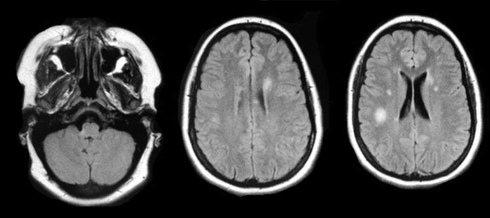
Brain mri scan showing multiple sclerosis lesions. Widespread use of mri (magnetic resonance imaging) has revolutionized the ability to diagnose multiple sclerosis. 39 calabrese m, filippi m, rovaris m et al. In fact, researchers and medical professionals consider mri to be one of the biggest breakthroughs in the field of multiple sclerosis, since it makes it possible to see lesions on the brain and spinal cord. Multiple sclerosis and mri 3.05.19 although the cause of multiple sclerosis (ms) is unknown ongoing research is moving forward at a remarkable pace and more potential therapies appear to be in the pipeline than ever before.
Ms Composite from case.edu An mri scan is abnormal in more than 95% of people recently diagnosed with ms. Abnormalities show up on scans from many illnesses other than ms. Multiple sclerosis and mri 3.05.19 although the cause of multiple sclerosis (ms) is unknown ongoing research is moving forward at a remarkable pace and more potential therapies appear to be in the pipeline than ever before. But abnormal mri results do not always mean that you have ms. In fact, researchers and medical professionals consider mri to be one of the biggest breakthroughs in the field of multiple sclerosis, since it makes it possible to see lesions on the brain and spinal cord. Mri is an important diagnostic tool for multiple sclerosis because it produces images of lesions in the brain and spinal cord. In ms, the immune system attacks the protective sheath (myelin) that covers nerve fibers and causes communication problems between your brain and the rest of your body. Magnetic resonance imaging (mri) is one of the most important and most commonly used tools for diagnosing and monitoring multiple sclerosis (ms).
Magnetic resonance imaging (mri) is one of the most important and most commonly used tools for diagnosing and monitoring multiple sclerosis (ms).
Abnormalities show up on scans from many illnesses other than ms. A multiple sclerosis diagnosis may not always be made solely on the basis of mri. An mri scan is the best way to locate multiple sclerosis (ms) lesions (also called plaques) in the brain or spinal cord. Magnetic resonance imaging (mri) has played a central role in the clinical management and scientific investigation of multiple sclerosis (ms) and has become the most important ancillary tool for diagnosing and monitoring the disease. The clinical usage of … In fact, researchers and medical professionals consider mri to be one of the biggest breakthroughs in the field of multiple sclerosis, since it makes it possible to see lesions on the brain and spinal cord. Sponsored by the consortium of ms centers, an international group of neurologists, radiologists, and imaging. Multiple sclerosis (ms) is a relatively common acquired chronic relapsing demyelinating disease involving the central nervous system, and is the second most common cause of neurological impairment in young adults, after trauma 19. Since its technical development in the early 1980s, magnetic resonance imaging (mri) has quickly been adopted as an essential tool in supporting the diagnosis, longitudinal monitoring, evaluation of therapeutic response, and scientific investigations in multiple sclerosis (ms). Mri is an important diagnostic tool for multiple sclerosis because it produces images of lesions in the brain and spinal cord. These lesions can appear as the condition progresses, and they may. The diagnosis is made from a combination of clinical, imaging, and laboratory findings patients with ms can present with motor, sensory, visual, and/or autonomic pathway symptoms Magnetic resonance imaging (mri) is one of the most important and most commonly used tools for diagnosing and monitoring multiple sclerosis (ms).
Morphology and evolution of cortical lesions in multiple sclerosis. But abnormal mri results do not always mean that you have ms. 2017 (october) cmsc proposed mri protocol pdf (414.72 kb) more hide administration. In fact, researchers and medical professionals consider mri to be one of the biggest breakthroughs in the field of multiple sclerosis, since it makes it possible to see lesions on the brain and spinal cord. A multiple sclerosis diagnosis may not always be made solely on the basis of mri.
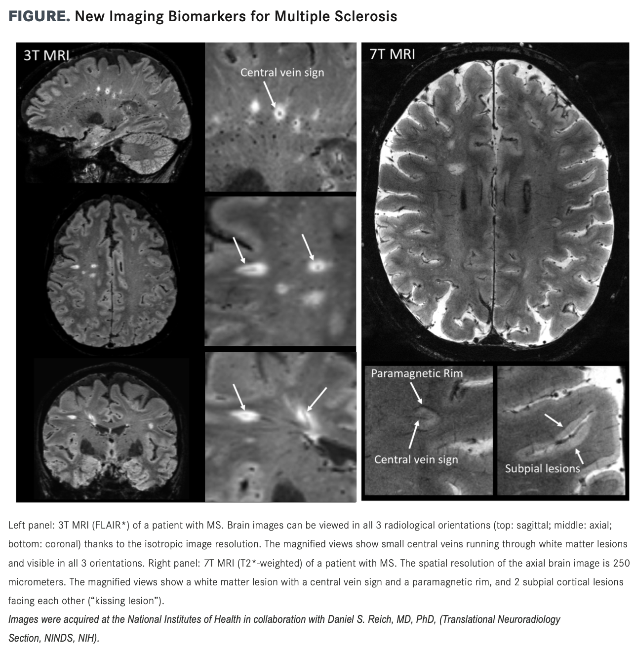
The Future Of Multiple Sclerosis Imaging from cdn.sanity.io Since its technical development in the early 1980s, magnetic resonance imaging (mri) has quickly been adopted as an essential tool in supporting the diagnosis, longitudinal monitoring, evaluation of therapeutic response, and scientific investigations in multiple sclerosis (ms). Morphology and evolution of cortical lesions in multiple sclerosis. Magnetic resonance imaging (mri) is one of the most important and most commonly used tools for diagnosing and monitoring multiple sclerosis (ms). Magnetic resonance imaging (mri) plays a crucial role in multiple sclerosis (ms) diagnosis, disease monitoring, prognostication, and research. The clinical usage of … Brain mri scan showing multiple sclerosis lesions. An mri scan is the best way to locate multiple sclerosis (ms) lesions (also called plaques) in the brain or spinal cord. Magnetic resonance imaging (mri) has developed into the most important tool for the diagnosis and monitoring of multiple sclerosis (ms).
For the first time, experts in multiple sclerosis (ms) from north america and europe have aligned on consensus recommendations for the use of mri in people with ms.
Widespread use of mri (magnetic resonance imaging) has revolutionized the ability to diagnose multiple sclerosis. Its high sensitivity for the evaluation of inflammatory and neurodegenerative processes in the brain and spinal cord has made it the most commonly used technique for the evaluation of patients with ms. The clinical usage of … The diagnosis is made from a combination of clinical, imaging, and laboratory findings patients with ms can present with motor, sensory, visual, and/or autonomic pathway symptoms Magnetic resonance imaging (mri) is the gold standard imaging technique for the identification of demyelinating lesions which can be used to support a clinical diagnosis of ms, and ms can now be diagnosed in some patients after a clinically isolated syndrome (cis) using new mri diagnostic criteria. In fact, researchers and medical professionals consider mri to be one of the biggest breakthroughs in the field of multiple sclerosis, since it makes it possible to see lesions on the brain and spinal cord. Sponsored by the consortium of ms centers, an international group of neurologists, radiologists, and imaging. Morphology and evolution of cortical lesions in multiple sclerosis. It is the preferred imaging method to help establish a diagnosis of ms and to monitor the course of the disease. Magnetic resonance imaging (mri) has developed into the most important tool for the diagnosis and monitoring of multiple sclerosis (ms). Mri and ms multiple sclerosis (ms) is a condition in which the body's immune system attacks the protective covering (myelin) surrounding the nerves of the central nervous system (cns). Multiple sclerosis multiple sclerosis (ms) is a potentially disabling disease of the brain and spinal cord (central nervous system). An mri scan is the best way to locate multiple sclerosis (ms) lesions (also called plaques) in the brain or spinal cord.
Brain mri scan showing multiple sclerosis lesions multiple sclerosis. Multiple sclerosis and mri 3.05.19 although the cause of multiple sclerosis (ms) is unknown ongoing research is moving forward at a remarkable pace and more potential therapies appear to be in the pipeline than ever before.